

Cryonics has exactly one irreversible chemistry problem to solve, and this is the phase where it gets solved. The patient's water has to be replaced with a medical antifreeze, evenly, before the body is cooled, because water that stays behind will freeze into crystals and crystals destroy the very structures we are trying to keep. The first-response team bought the time. The surgical phase is where that time is spent buying the only thing that makes long-term storage worthwhile: a body that can turn to glass instead of ice.
This is real surgery with real plumbing, performed in a specialized field unit equipped for cryoprotective perfusion. It is worth walking through it concretely, because the details are where preservation quality lives.
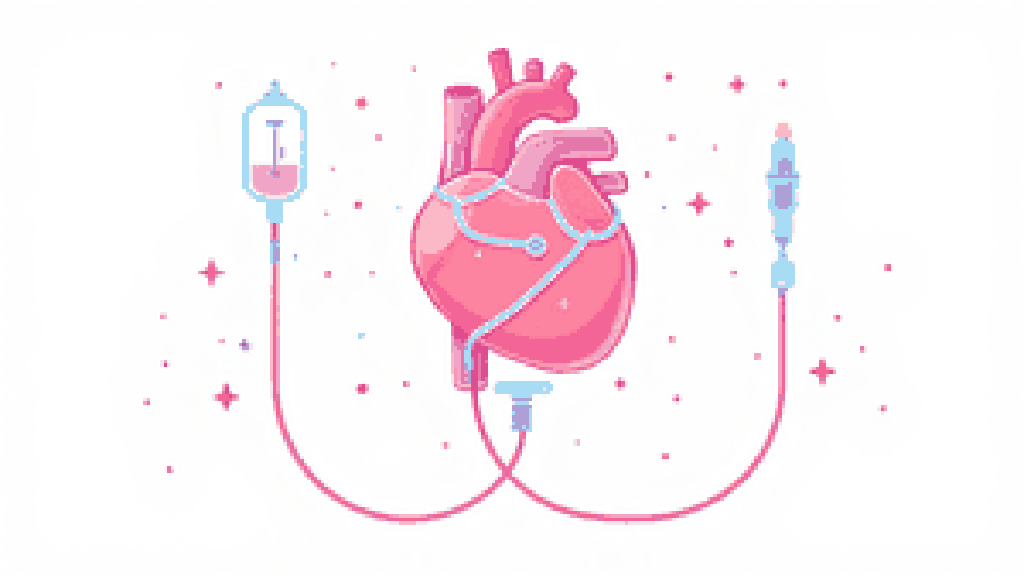
Glass, not ice: what the surgery is actually for
The entire procedure exists to enable vitrification. Ordinary freezing forms ice, and ice expands and shreds cell membranes. The way around it is to perfuse the body with cryoprotective agents (CPAs) that replace much of the body's water, so that when the tissue is cooled it sets into a glass with no crystals at all. That is the difference between preserving a structure and destroying it, and it is also why this phase is distinct from chemical fixation: the goal is a vitrifiable patient, not a chemically frozen one.
Setup: reading the clock the first team left behind
Before anyone makes an incision, the surgical team reviews the first-response record: the time of legal death, the cooling profile, the stabilization timeline, and how much ischemic exposure the patient has already had. That history is not bureaucracy; it tells the team what they are working with and how aggressive the perfusion needs to be. The patient's temperature should already be within the protocol's range, having been cooled hard during first response and stabilization, and vascular access is prepared.
The procedure, step by step
The perfusion itself is a controlled, monitored sequence, not a single push of fluid:
- Cannulation. Major vessels are selected and accessed, and a closed perfusion circuit is established with flow and pressure monitoring and waste collection. Closed, because every variable has to be watched.
- Initial flush. The blood is replaced with a base perfusate. This clears clots, washes out metabolic waste, and stabilizes fluid volume, so the body is a clean canvas before the antifreeze goes in.
- Cryoprotectant ramp. The CPA is introduced in a graded sequence, rising in concentration step by step rather than all at once. This is the most delicate part: jump the concentration too fast and the cells suffer osmotic shock, as water is yanked out of them too quickly. The perfusion temperature is usually lowered gradually in parallel, because CPAs are mildly toxic and that toxicity drops as the tissue gets colder.
- Temperature and flow monitoring. Throughout, the team holds target flow rates and pressures matched to vessel size and condition, and watches for uniform cooling, for the CPA concentration at the outlet rising to meet the inlet, and for changes in vascular resistance that signal trouble.
- Endpoint. Perfusion stops when the effluent shows that enough CPA has saturated the tissue and the vasculature has equilibrated. Access points are sealed and the patient is readied for controlled cooldown.
Where it can go wrong, honestly
Calibration over reassurance: this phase is good, not perfect, and the failure modes are worth naming. CPA toxicity rises with temperature, which is exactly why perfusion is done cold, accepting some toxicity as the price of avoiding ice. Vascular integrity is the other constraint that bites: perfusion can only reach tissue the blood vessels can carry it to, so any region the circuit cannot supply, because of clots, damage, or prior ischemia, stays under-perfused and is where ice damage shows up later. This is one of the central technical challenges for high-quality preservation, and real-time monitoring exists precisely to catch unevenness while it can still be corrected.
The honest version of the bet is this: perfusion will not be perfectly even, and the cryoprotectant is not perfectly benign, but the wager is that the damage spares the information in the brain's structure even where it stresses the tissue, and that future repair can work from a preserved structure. That is the same expected-value reasoning behind biostasis as a whole, applied to a surgical table.
The outcome
At the end of the procedure, the patient's tissues, above all the neural tissue, are saturated with vitrification-capable solution, fluid replacement is complete, and the patient is ready for controlled cooldown toward storage temperature. From here the path runs to the long-term storage facility, where the glass that this surgery made possible is held stable, in principle, indefinitely.
This surgery is the moment a body stops being something that will rot and becomes something that can turn to glass; everything before it is preparation and everything after it is patience.